


 Author: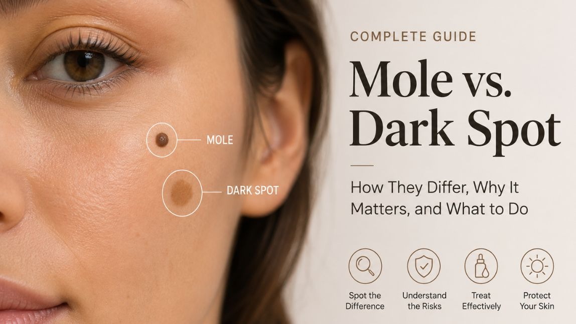
Reviewed by the Perfect Corp. Skin AI research team. This article is educational and is not a substitute for a clinical skin examination.
Two brown marks on the same cheek. One has been there since you were nine. The other showed up after a summer in Lisbon. They look almost identical in the bathroom mirror, and yet they are biologically different structures that behave differently, respond to completely different treatments, and carry very different risk profiles.
Getting the distinction right matters more than most people assume. It changes whether you reach for a vitamin C serum or book a dermatology appointment. It also explains a question we get constantly from brands deploying skin analysis technology: why does the AI score my dark spots but seem to ignore my moles?
Mole vs. Dark Spot: The Short Answer
A mole is a growth made of clustered pigment cells (melanocytes) that sit deeper in the skin, so it often has height and texture you can feel. A dark spot is flat pigment sitting in the upper layers of the skin, caused by melanin overproduction from sun, hormones, or past inflammation. Run a fingertip over it: moles usually have relief, dark spots do not.
That fingertip test resolves maybe 80% of cases. The rest need a closer look, which is what the rest of this article is for.
What Is a Mole?
The medical term is a nevus (plural: nevi). Moles form when melanocytes, the cells that manufacture your skin's pigment, grow in a cluster instead of distributing evenly across the skin. That clustering is the entire distinction. Same cell type as the rest of your skin, different architecture.
What that architecture produces:
- Defined, even borders. A benign mole tends to have a crisp edge where it stops and normal skin begins.
- Consistent color throughout. Usually one shade of tan, brown, or black, though moles can also be flesh-toned, pink, blue, or red.
- Round or oval shape. Symmetrical enough that folding it in half mentally gives you two matching halves.
- Height, in many cases. Raised, dome-shaped, or slightly wrinkled. Some are perfectly flat, which is where confusion starts.
- Occasional hair. Dark coarse hair growing from a mole is generally a reassuring sign, not an alarming one.
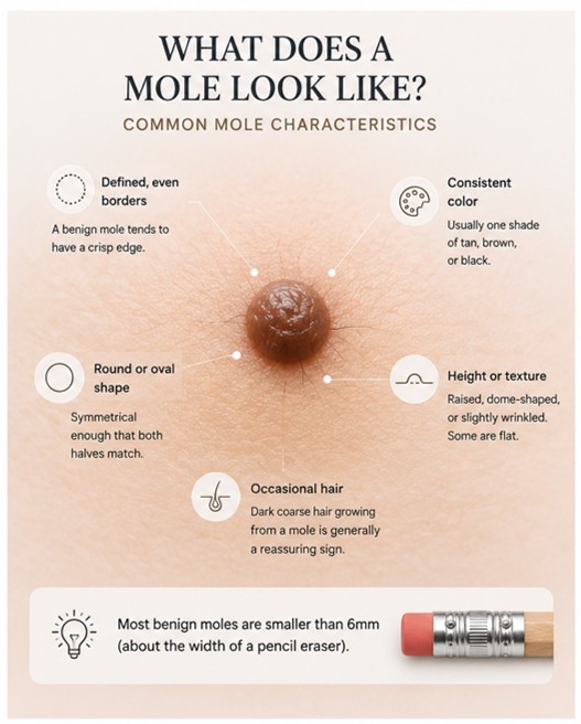
Most people carry somewhere between 10 and 40 moles by adulthood. They typically appear during childhood and adolescence, keep arriving until roughly age 40, and then some begin to fade or disappear entirely. New moles after 40 are worth mentioning to a dermatologist simply because they are off-schedule.
Size is a useful benchmark: most benign moles measure under 6mm across, roughly the width of a pencil eraser.
What Is a Dark Spot?
"Dark spot" is not one condition. It is a shorthand for several types of hyperpigmentation, all sharing one mechanism: melanocytes producing too much melanin in response to a trigger, then depositing it into the surrounding skin. The pigment sits mostly in the epidermis, which is why these marks stay flat and why topical treatment can reach them.
The four types you are most likely dealing with
- Solar lentigines (sun spots, age spots). Flat, tan to dark brown, usually larger than a freckle and with a slightly irregular outline. They appear on cumulative sun exposure areas: cheeks, hands, forearms, chest. They do not fade in winter.
- Ephelides (freckles). Small, light brown, and genetically driven, often tied to the MC1R gene and fair skin. They darken in summer and lighten when sun exposure drops, which is the single easiest way to tell them apart from sun spots.
- Melasma. Larger, patchy, often roughly symmetrical across the forehead, upper lip, and cheekbones. Hormonally triggered by pregnancy, oral contraceptives, or thyroid changes, and aggravated by both UV and visible light.
- Post-inflammatory hyperpigmentation (PIH). The mark left behind after acne, eczema, a burn, or a bug bite heals. It traces the shape of whatever caused it, which is a strong diagnostic clue.
All four are cosmetic concerns rather than medical ones. That does not make them trivial to a consumer, and it is precisely why pigmentation is one of the highest-intent categories in skincare retail.
Mole vs. Dark Spot: Key Differences at a Glance
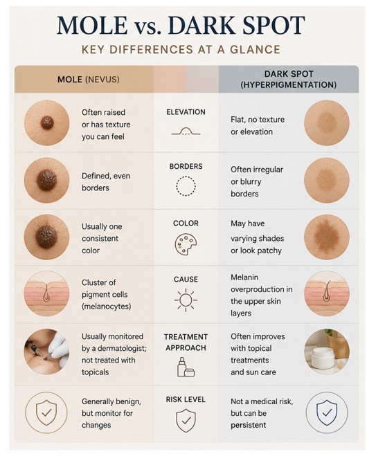
| Feature | Mole (Nevus) | Dark Spot (Hyperpigmentation) |
|---|---|---|
| What it is | A growth of clustered melanocytes | Excess melanin deposited in normal skin |
| Depth | Extends into the dermis | Mostly epidermal, sits near the surface |
| Texture | Often raised, you can feel it | Flat, level with surrounding skin |
| Border | Sharply defined, usually round or oval | Softer, more diffuse, sometimes patchy |
| Cause | Genetics, with sun and hormones as amplifiers | UV exposure, hormones, or past inflammation |
| Typical onset | Childhood through early adulthood | Any age, usually after the triggering event |
| Seasonal change | Stable year-round | Freckles fade in winter, sun spots do not |
| Response to skincare | Does not fade with topicals | Often responds to tyrosinase inhibitors, retinoids, exfoliation |
| Removal | Clinical procedure only | Topicals, chemical peels, or laser |
| Cancer relevance | Melanoma can arise from or resemble a mole | Low risk, but lentigo maligna can imitate a sun spot |
The Four-Question Check You Can Run at Home
Dermatologists work from pattern recognition built over thousands of cases. You can borrow a simplified version of the same logic. Ask four questions in order.
1. Can you feel it?
Close your eyes and run a fingertip across the mark, then across the skin beside it. Any bump, any change in drag, any texture at all points toward a mole or a seborrheic keratosis. Perfectly smooth points toward pigmentation.
2. When did it arrive?
Marks you have had since childhood are almost always moles or freckles. Marks that appeared in your thirties or later, on sun-exposed skin, are almost always sun spots. Marks that appeared exactly where a pimple was two months ago are PIH.
3. Does it change with the seasons?
Freckles darken in August and lighten by February. Sun spots and moles hold steady. If a mark has a summer personality and a winter personality, it is pigment responding to UV, not a growth.
4. Is it symmetrical, and is it stable?
Draw an imaginary line through the middle. Benign moles match on both sides. Then compare against a photo from six months ago. Stability is the single most reassuring signal in all of skin assessment, and change is the single most important one to act on.
Mole vs. Freckle vs. Sun Spot vs. Seborrheic Keratosis
Most articles stop at moles and freckles. In practice, six things routinely get confused with each other, and two of them are frequently misfiled as "dark spots" when they are actually growths.
| Type | Texture | Color | Trigger | Fades on its own? |
|---|---|---|---|---|
| Mole (nevus) | Flat or raised | Tan to black, uniform | Genetic | Sometimes, over decades |
| Freckle (ephelide) | Flat | Light brown | Genetic plus UV | Yes, seasonally |
| Sun spot (solar lentigo) | Flat | Tan to dark brown | Cumulative UV | No |
| Melasma | Flat | Brown to grey-brown patches | Hormones plus light | Rarely, often recurs |
| PIH | Flat | Pink, brown, or grey | Prior inflammation | Yes, over months |
| Seborrheic keratosis | Raised, waxy, "stuck on" | Tan to dark brown or black | Age and genetics | No |
Seborrheic keratosis deserves a flag. It is benign, extremely common after 40, and looks like someone pressed a piece of dark wax onto the skin. People assume it is a suspicious mole and panic, or assume it is a stubborn dark spot and spend a year on brightening serums that were never going to work.
Do Moles and Dark Spots Look Different on Deeper Skin Tones?
Yes, and this is where most consumer-facing content quietly fails.
On Fitzpatrick types IV through VI, the contrast between a pigmented mark and the surrounding skin is lower, so both moles and hyperpigmentation can be harder to spot early. Two practical consequences:
- PIH is more common and more persistent. Deeper skin tones produce a stronger pigment response to inflammation, which is why acne marks can outlast the acne by many months.
- Melanoma more often appears in low-UV areas. On darker skin, it disproportionately shows up on palms, soles, under nails, and inside the mouth. A dark streak under a fingernail or a changing mark on the sole of a foot warrants a professional look, regardless of how much sun that area has seen.
This is also a real technical requirement for any brand deploying diagnostic tech. A pigmentation model trained mostly on light skin will under-detect on deep skin, which turns a personalization feature into a trust problem. Perfect Corp.'s skin models are trained on a dataset of over 70,000 medical-grade images spanning diverse tones, types, and textures, specifically so scoring holds up across the full Fitzpatrick range. You can see how that plays out in practice in our breakdown of AI face mapping for skin analysis.
The ABCDE Rule: When a Mole Needs a Dermatologist
This is the standard dermatology screening mnemonic, and it is worth memorizing properly rather than half-remembering it.
- A is for Asymmetry. One half does not mirror the other.
- B is for Border. Edges are ragged, notched, blurred, or bleeding into surrounding skin.
- C is for Color. More than one shade within a single mark, or a color that is unusual for you.
- D is for Diameter. Larger than about 6mm, though melanoma can be smaller.
- E is for Evolving. Any change in size, shape, color, or elevation, or new itching, crusting, or bleeding.
Two additions worth knowing. The "ugly duckling" sign says that a mark that looks meaningfully different from every other mole on your body deserves attention even if it passes ABCDE. And E carries the most weight of the five. A mole that has looked odd for twenty years is far less concerning than one that changed last month.
Can a Dark Spot Turn Into Melanoma?
A sun spot does not transform into melanoma the way a caterpillar becomes a butterfly. That is not the mechanism.
The real concern is different and more subtle: lentigo maligna, an early form of melanoma, can look almost exactly like an ordinary sun spot in its first years. It is typically a slowly enlarging flat brown patch on chronically sun-exposed skin, most often the face, in people over 60. It expands and develops uneven color over time rather than staying put.
Separately, having a lot of sun spots is a marker that your skin has absorbed significant lifetime UV, which independently raises skin cancer risk in those areas. The spots are the receipt, not the disease.
Practical rule: a flat brown patch that keeps growing or keeps changing color needs a professional opinion, whatever you have been calling it.
Can AI Skin Analysis Detect Moles?
Here is where we have to be precise, because the answer is "it depends what you mean by detect."
Consumer and retail AI skin analysis, the kind that powers a brand's skincare recommendation experience, is built to assess cosmetic skin condition. It measures things like spots, wrinkles, pores, texture, redness, oiliness, moisture, firmness, radiance, dark circles, eye bags, and acne, then scores each one and maps it to a region of the face. Perfect Corp.'s system evaluates 15 concerns and 8 skin types in a single scan, with 95% test-retest reliability and over 80% correlation with physician evaluation in an independent study.
What it does not do, and is not designed to do, is triage moles for malignancy. That is a regulated medical device category with an entirely different validation pathway, a different training corpus, and a different risk calculus. Dermoscopy-based lesion classification tools exist and are genuinely useful, but they are separate products with clinical clearance behind them.
So: a cosmetic skin analysis engine can see a mole in the image. It will not tell you whether it is dangerous, and any tool that implies otherwise without clearance should be treated with suspicion.
Why Moles Are Excluded From AI Skin Analysis Spot Scores
This is the part most people find counterintuitive. If the AI can see the mole, why does it not count it in the spot score?
Because counting it would make the score wrong.
The "spots" metric in a skin analysis report is answering a specific question: how much treatable pigmentation is present, and is it improving? That number needs to move when a customer starts using a brightening serum. A mole will not move. It is a permanent structure, not a pigmentation state.
Three concrete reasons pigment models are trained to separate them:
- Score integrity over time. If moles inflate the spot count, a customer with twelve facial moles starts at an artificially poor baseline and never improves, no matter how well their routine works. Progress tracking breaks.
- Recommendation accuracy. A high spot score triggers pigmentation product recommendations. Recommending niacinamide because the model counted three moles is a bad recommendation, and customers notice.
- Staying inside the cosmetic lane. Scoring moles as a "concern" implies they are a problem to be fixed, which edges a beauty tool toward medical claims it is not licensed to make.
Well-built models handle this through morphological separation. Moles present as compact, high-contrast, sharply bounded, texturally distinct regions. Hyperpigmentation presents as diffuse, lower-contrast, soft-edged areas within otherwise normal skin. Those signatures are different enough that a model trained on labeled dermatological data can reliably route each into its own bucket. We go deeper into how these detection pipelines are architected in our guide to how AI skin analysis machines work.
How AI Skin Analysis Identifies Dark Spots
The pigmentation pipeline runs roughly like this:
- Capture and normalize. The system corrects for lighting temperature and exposure. Uncontrolled lighting is the single biggest source of noise in consumer selfie analysis, so normalization happens before anything else.
- Facial region segmentation. The face is divided into zones (forehead, cheeks, nose, chin, periorbital) so results are spatially specific rather than one global number. Perfect Corp. uses 180 degree mapping across front, left, and right captures, since lateral cheek pigmentation is frequently invisible head-on.
- Melanin channel isolation. Pigment signal is separated from redness signal, which prevents post-acne erythema from being scored as brown pigmentation.
- Lesion classification. Detected regions are sorted by contrast, edge gradient, size, and shape. This is the step where moles are separated out.
- Severity scoring and tracking. Each zone gets a numerical score, stored so the next scan can be compared against it.
That last step is the commercially interesting one. A single score is a curiosity. A score with a trend line is a reason to come back, and a reason to keep buying the product that is moving it. You can try the full flow yourself on the Perfect Corp. skincare demo store.
When Should You See a Dermatologist?
Book an appointment if any of the following apply:
- A mole meets any ABCDE criterion, especially E
- Any mark bleeds, itches persistently, crusts, or fails to heal
- A new mole appears after age 40
- One mole looks distinctly unlike all your others
- A flat brown patch keeps enlarging or developing uneven color
- A dark streak appears under a fingernail or toenail
- Many dark spots appear suddenly without an obvious sun or hormonal trigger
- You have a personal or family history of melanoma, over 50 moles, or significant sun damage
For everyone else, annual professional skin checks are a reasonable baseline, and monthly self-examination in good light costs five minutes.
Nothing in this article, and no consumer AI tool, replaces that examination. Skin analysis technology is for understanding and tracking cosmetic skin condition. Diagnosis belongs to a clinician.
Tracking Your Skin Over Time
Whether a mark is a mole or a dark spot, the thing that actually generates useful information is longitudinal comparison. Human memory is unreliable at exactly this task. Nobody can recall the precise border of a mark from four months ago.
Two habits close that gap. First, photograph anything you are watching in consistent lighting, with a ruler or coin for scale, at fixed intervals. Second, use structured scoring for the cosmetic side, so pigmentation improvement is measured rather than guessed at.
For beauty brands, this is the entire strategic argument for skin diagnostics. A customer who can see their spot score drop four points over eight weeks has a reason to trust the recommendation, repurchase the product, and return to the tool. Correctly excluding moles from that score is not a technical footnote. It is what keeps the number honest.
Explore AI Skin Analysis for Your Brand
See how accurate pigmentation detection, region-level scoring, and progress tracking work inside a real retail experience. Explore Perfect Corp.'s AI Skin Analysis solution or contact our team for a walkthrough tailored to your product catalog.
Frequently Asked Questions
How can you tell if it is a mole or a dark spot?
Touch it. Moles usually have height or texture because they are growths extending into deeper skin. Dark spots are flat pigment sitting near the surface. Then check timing: lifelong marks tend to be moles, marks that appeared later on sun-exposed skin tend to be sun spots.
Can a dark spot turn into a mole?
No. They are different structures. A dark spot is excess pigment in otherwise normal skin, while a mole is a cluster of pigment cells. Neither converts into the other.
Are moles and dark spots the same thing?
No. Both look brown, but a mole is a growth of clustered melanocytes with depth and defined borders, while a dark spot is surface-level hyperpigmentation caused by UV, hormones, or past inflammation. They also respond to completely different treatments.
Do dark spots fade on their own?
It depends on the type. Freckles lighten with reduced sun exposure. Post-inflammatory hyperpigmentation usually fades over several months. Sun spots and melasma generally do not resolve without treatment and consistent daily SPF.
Can moles be flat?
Yes. Junctional nevi are flat and can closely resemble a large freckle or sun spot. When texture does not settle the question, fall back on border definition, color uniformity, and how long the mark has been there.
Can AI skin analysis apps detect skin cancer?
Consumer beauty and retail skin analysis tools are built for cosmetic assessment and are not cleared to diagnose skin cancer. Separate dermoscopy-based medical devices exist for lesion triage. Treat any beauty app claiming cancer detection without regulatory clearance with caution.
Why does my skin analysis result not show my moles?
Because pigmentation models deliberately exclude them. Moles are permanent structures that will not respond to skincare, so counting them in a spot score would distort your baseline and make progress tracking meaningless.
What is the fastest way to fade a dark spot?
Daily broad-spectrum SPF is non-negotiable, since UV re-triggers pigment production faster than any active can reduce it. Beyond that, tyrosinase inhibitors such as vitamin C, niacinamide, azelaic acid, and tranexamic acid, plus retinoids for turnover. Expect eight to twelve weeks before visible change. In-clinic peels and lasers work faster but carry rebound risk on deeper skin tones.
Is a raised dark spot always a mole?
Not always. Seborrheic keratosis is a benign raised growth that looks waxy and stuck onto the skin, and it is very common after 40. Dermatofibromas and some warts can also present as raised pigmented bumps. If a raised mark is new or changing, get it examined.
How often should I check my skin?
A monthly self-check in bright light, using a mirror for your back and scalp, plus an annual professional exam. Increase frequency if you have over 50 moles, fair skin, a history of blistering sunburns, or family history of melanoma. You can also browse more skin-health and beauty tech guidance across the Perfect Corp. blog.



 English


